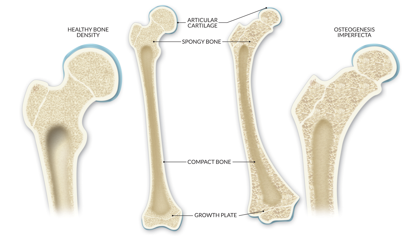
Osteogenesis Imperfecta (OI) is an inherited bone condition commonly known as “brittle bone disease” as it leads to bones that break more easily, from mild to extremely severe. 1
TL-HEX TrueLok Hexapod System™
Important safety information: Download the Product Instructions For Use.
TL-HEX is a dynamic, 3D external fixation system that combines hardware and software to correct bone deformities. This hexapod-based system functions as a 3D bone segment-repositioning module. In essence, the system consists of circular and semi-circular external supports secured to the bones by wires and half-pins, interconnected by six struts.
Important safety information: Download the Product Instructions For Use.
The Ilizarov System has experienced many modifications over the last fifty years. The TrueLok™ Ring Fixation System, developed at Texas Scottish Rite Hospital for Children (TSRHC) in Dallas, Texas, is one of the modern variants of the original fixator, but preserves many of the original principles of Professor Ilizarov. It consists of aluminum rings available in different sizes, connected to the bone through metal wires or/and bone screws. The relative movements of the rings allow the correction of almost all the bone deformities in upper and lower limbs and in the foot.
The underlying cause is usually a deficiency in the type or amount of collagen necessary for proper bone formation. It is most often inherited from a parent, but occasionally it may occur due to a chance alteration of the gene. As it is very rare, it is considered to be an orphan disease (0.005%). 2
Symptoms
The child may present with multiple or unusual fractures during childhood, or even before childbirth. There can be deformity within the bone, including curvature of the spine, and they may be smaller than average in height. The more severely affected may have breathing problems. There may be bone pain, and the teeth and hearing may be affected. Due to the abnormal collagen there may also be increased laxity of the ligaments, which can lead to joint problems, transparent skin and blue/grey sclerae (the white part of the eye). Some children may need to use a wheelchair to mobilize. 1
There are four main types of OI. The mildest form is Type I, characterized by very little bone deformity, but fragile bones and teeth, and blue sclerae of the eyes. Some patients with Type I OI are only mildly affected. Type II is a very severe form and the baby does not usually survive. Type III is the most severe survivable form, with fractures at birth that can be fatal, and very short stature. Type IV is of moderate severity: fractures and bowing of the bones are frequent but the sclerae and hearing are normal.
There are further rare types of OI.
Diagnosis
The diagnosis may be easy in a child with a family history of OI. It should also be considered in children with frequent fractures, especially following low impact trauma. The doctor will carefully examine the child’s skin, skull, bone alignment, teeth development and eyes to look for the signs of the condition.
X-rays can reveal new or previous fractures as well as deformity of the bones. A geneticist may perform blood or skin tests to find defective genes. A bone biopsy may detect a decrease in the bone volume and an increase in bone remodeling. 3 The most severe forms may be diagnosed on ultrasound scans during pregnancy.
While there is no cure for OI, there are many ways to improve a child’s quality of life. Medicationincluding bisphosphonates and vitamin D may improve the quality of the bone, prevent fractures and decrease pain.
Fractures may be treated with casts, slings, splints and braces if the bone is in good alignment. Movement and weight bearing is encouraged as soon as the bone has healed, according to the doctor’s evaluation. 4
If there is bone deformity due to fractured or weakened bone, surgery may be considered. This may be at the time of the fracture or a planned procedure. Fixation inside the bone, with rods or nails, may be used and will further improve the strength of the bone. Telescopic rods that can grow with the child may be used. External fixation may also be used. 5
Soft tissue surgery may be required to release contractures, such as the Achilles tendon, for example.
Severe scoliosis may require spinal fusion surgery as the fragile ribs may not allow bracing.
Throughout childhood, regular hospital specialist consultations and input from allied health care professionals, including physiotherapyand occupational therapists, are needed.
Complications
Following surgery with telescopic rods, further surgery may be required as the rods may move within the soft bone, and sometimes perforate the joint or soft tissues. Infections and pseudoarthrosis when the bone does not heal are other common complications.
OI tends to have less effect in young adult life, as falls and injuries are less frequent. However, bone fragility and fractures increase again in older age, especially after menopause for women. To maintain bone health, patients must be advised not to smoke and to refrain from drinking alcohol.
Genetic Home Reference 2020. US National Library of Medicine. Osteogenesis Imperfecta.
Pediatric Orthopaedic Society of North America (POSNA). Orthogenesis Imperfecta.
Souder C. 2019. Osteogenesis Imperfecta.
American Academy of Orthopaedic Surgeons (AAOS). Osteogenesis Imperfecta.
Popkov D. 2018. Use of intramedullary nailing in combination with external fixator for a postoperative defect and pseudoarthrosis of femur in a girl with osteogenesis imperfecta type VIII: a case report. Strategies Trauma Limb Reconstr. 13(3):191-7.