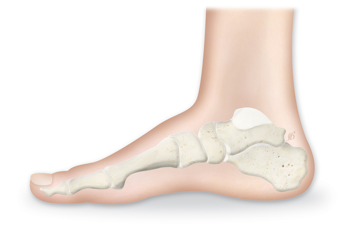
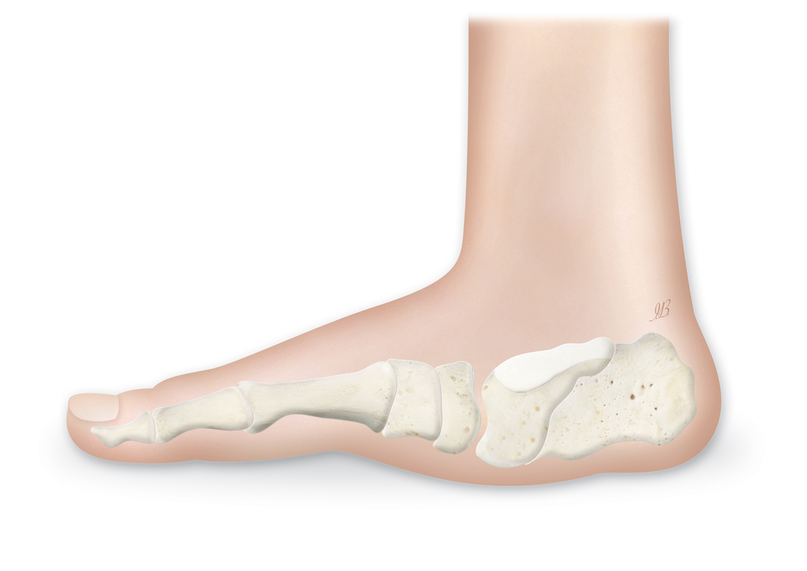
Diabetic Charcot foot is a chronic and progressive (it gets worse over time) joint disease following the development of neuropathy (the loss of protective sensation in the limb); it occurs in about 1 percent of neuropathic patients.1
Normal Foot
Charcot Foot
Ankle Hindfoot Nailing System™
Important safety information: Download the Product Instructions For Use.
The Orthofix Ankle Hindfoot Nailing System (AHN) is designed to be used for the fusing and fixation of bones in the ankle.
Important safety information: Download the Product Instructions For Use.
In beaming, a large diameter cannulated device distributes the axial load of the bone between fixation points, which helps hold a joint or osteotomy until it heals.
Important safety information: Download the Product Instructions For Use.
TL-HEX is a dynamic, 3D external fixation system that combines hardware and software to correct bone deformities. This hexapod-based system functions as a 3D bone segment-repositioning module. In essence, the system consists of circular and semi-circular external supports secured to the bones by wires and half-pins, interconnected by six struts.
Important safety information: Download the Product Instructions For Use.
The Ilizarov System has experienced many modifications over the last fifty years. The TrueLok™ Ring Fixation System, developed at Texas Scottish Rite Hospital for Children (TSRHC) in Dallas, Texas, is one of the modern variants of the original fixator, but preserves many of the original principles of Professor Ilizarov. It consists of aluminum rings available in different sizes, connected to the bone through metal wires or/and bone screws. The relative movements of the rings allow the correction of almost all the bone deformities in upper and lower limbs and in the foot.
Charcot foot had been documented to occur as a consequence of various peripheral neuropathies (lessened ability to feel sensations in the limbs); however, diabetic neuropathy has now become the most common cause. The interaction of several factors (diabetes, sensory-motor neuropathy, autonomic neuropathy, trauma, and metabolic abnormalities of bone) results in acute, local inflammation, which may lead to varying degrees and patterns of bone destruction, dislocation and deformity.1
Symptoms
The hallmark deformity associated with this condition is midfoot collapse, described as a “rocker-bottom” foot, although the condition appears in other joints and with other presentations. Pain or discomfort may be a feature of this disorder at the active (acute) stage, but the level of pain may be significantly diminished when compared with individuals with normal sensation and equivalent degrees of injury.1
Diagnosis
An X-ray will confirm a diagnosis of Charcot foot.
Charcot foot can be managed with physical therapy, braces or casting, pain medicationand surgery. For severe deformities that cannot be treated in a brace, osteotomy(the surgical cutting of a bone or removal of a piece of bone) and some sort of externaland/or internal fixation device can help to reverse foot and joint deformities, aiding support and strength. Arthrodesis(fusion of the joints) and amputation are other treatment options, depending on the severity of the condition.
Complications
Left untreated, Charcot foot leads to pain and difficulties walking, which decreases everyday activity levels and, in turn, adversely affects quality of life with further deteriorating mobility. Foot wounds, foot fractures and more severe deformities of the foot and ankle may also occur. It is important to remember that the underlying conditions, such as diabetes, should be managed over the long-term, even after Charcot foot is treated in the “active” stage.