Home / Limb Deformities / Clubfoot
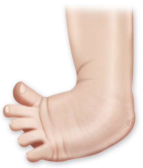
Clubfoot is diagnosed when a baby’s foot is twisted down and in. Usually present at birth, clubfoot occurs in about 1.3 per 1,000 births (0.13 percent), and is usually an isolated problem for an otherwise-healthy baby.1 About half of children with clubfoot have the condition in both feet.
Important safety information: Download the Product Instructions For Use.
The Guided Growth System eight-Plate and quad-Plate is an implant system designed to correct pediatric deformities (congenital or acquired) in lower limb, like genu valgum and varum, and limb length discrepancies. The plates are attached to the external surface of the bone over the growth plate by two or four screws depending on the plate selected by the surgeon. The implant acts like a flexible hinge, permitting growth at the growth plate to gradually straighten the limb when the physis (growth plates) are not fused. The screws are not locked to the plate but rather are allowed to swivel and diverge in their positions as the bone grows. Immediately after implantation, the patient is allowed mobility and weight bearing.
Important safety information: Download the Product Instructions For Use.
The eight-Plate Guided Growth System+™ is an implant system designed to correct pediatric deformities (congenital or acquired) in lower limb, like genu valgum and varum, and limb length discrepancies. The plates are attached to the external surface of the bone over the growth plate by two or four screws depending on the plate selected by the surgeon. The implant acts like a flexible hinge, permitting growth at the growth plate to gradually straighten the limb when the physis (growth plates) are not fused. The screws are not locked to the plate but rather are allowed to swivel and diverge in their positions as the bone grows. Immediately after implantation, the patient is allowed mobility and weight bearing. The eight-Plate Guided Growth System+ offers more sizes and different screw types compared to the standard eight-Plate Guided Growth System, as well as an increased screw angle to treat very difficult cases.
Important safety information: Download the Product Instructions For Use.
TL-HEX is a dynamic, 3D external fixation system that combines hardware and software to correct bone deformities. This hexapod-based system functions as a 3D bone segment-repositioning module. In essence, the system consists of circular and semi-circular external supports secured to the bones by wires and half-pins, interconnected by six struts.
Important safety information: Download the Product Instructions For Use.
The Ilizarov System has experienced many modifications over the last fifty years. The TrueLok™ Ring Fixation System, developed at Texas Scottish Rite Hospital for Children (TSRHC) in Dallas, Texas, is one of the modern variants of the original fixator, but preserves many of the original principles of Professor Ilizarov. It consists of aluminum rings available in different sizes, connected to the bone through metal wires or/and bone screws. The relative movements of the rings allow the correction of almost all the bone deformities in upper and lower limbs and in the foot.
Important safety information: Download the Product Instructions For Use.
The MiniRail External Fixation System consists of small rails and screws for fixation of small bones when a condition does not allow for implanting internal devices. The MiniRail is designed to help with a variety of deformity corrections and lengthening procedures of small bones and joints in the foot and upper limb.

The cause is at least partly genetic because it is known to run in families. Otherwise, the cause is not known, but it might happen when muscles are not even in the lower leg.
In clubfoot, the top of the foot usually twists down and in. The arch is high, and the heel also turns in. In more-severe cases, the foot might actually look upside-down. Even though clubfoot may shorten the foot and affect normal growth of the calf muscles, clubfoot itself doesn’t actually cause discomfort or pain.

A doctor can usually diagnose clubfoot based on how the foot looks after birth or sometimes also during routine ultrasound scans during pregnancy. In some cases, X-rays may be needed to determine the severity of the condition.

Clubfoot hinders normal walking so treatment should begin soon after birth. The most common treatment method involves stretching and casting (to hold the new position of the foot), performed repeatedly over several months, followed by minor surgery to lengthen the Achilles tendon under local anesthetic (Ponseti method). Stretching and use of special shoes and braces may be needed for up to three years after the surgery. Most children treated this way will have pain-free, normal-looking feet that function well.
However, severe clubfoot that has not gotten better with stretching and casting may need surgery and possibly the use of external fixation, which can be used to help reshape the muscles and other soft tissue over the course of several months to one year. 2 In most cases, babies who are treated early are able to wear ordinary shoes and participate in normal activities when they are older.

Even after treatment, once your child starts to stand and walk, mobility may be slightly limited. In addition, shoe sizes may differ between the clubfoot and the unaffected foot. If left untreated, your child is likely to have a lack of normal muscle growth, arthritis, inability to walk normally and, perhaps, self-esteem issues related to how the foot looks.